Right Leg Bone Diagram - Joints Ligaments And Connective Tissues Advanced Anatomy 2nd Ed. In the leg muscles diagram above, there are many muscles that make up your legs and support it to move. The femur, or thighbone, is the longest and largest bone in the human body. The tibia is the main bone of the leg, forming what is more commonly known as the shin. Normal leg bones are relatively straight, but those affected by paget's disease are porous and curved. While some people with paget's disease have no symptoms, others figure 9.
The sensory and motor innervation to the lower limb is supplied by the lumbosacral plexus, which is formed by the ventral rami of the lumbar and sacral spinal nerves with additional nerves of the right lower extremity, posterior view. In the centre of your chest. Its lower end helps create the knee joint. In the lower leg, the tibia bears most of the body's weight while the fibula supports the muscles of balance in the lower leg and ankle. Hand bone anatomy, wrist anatomy, anatomy bones, human body anatomy, human anatomy and physiology, muscle anatomy, musculoskeletal system, anatomy study, anatomy reference.
The Right Tibia And Fibula 6 Download Scientific Diagram from www.researchgate.net Start learning with our skeleton diagrams, bone labeling exercises and skeletal system quizzes! Can i also check that what you really require by arm and leg bone. Body weight is distributed among the seven tarsals, which can shift slightly to provide minute. The foot bones shown in this diagram are the talus, navicular, cuneiform, cuboid, metatarsals and calcaneus. Bones of the right leg. Reader view compact bone spongy bone of the long arm and leg bones that makes new red blood cells While some people with paget's disease have no symptoms, others figure 9. Molly smith dipcnm, mbant • reviewer:
Normal leg bones are relatively straight, but those affected by paget's disease are porous and curved.
Diagram of blood and nerve supply to bone. Lower jaw (mandible) collar bone. The bones of the lower extremity in man and of any of the four extremities in animals. Equivalent to leg bone and in_right_side_of some appendicular skeleton. The sensory and motor innervation to the lower limb is supplied by the lumbosacral plexus, which is formed by the ventral rami of the lumbar and sacral spinal nerves with additional nerves of the right lower extremity, posterior view. Although an inferred awareness of. Mesh obo (open biomedical ontologies). The tibia is the main bone of the leg, forming what is more commonly known as the shin. Learn the bones of the body with skeletal system quizzes. If encode really needs these we will add them. Bones of the pelvic girdle. The admission, the patient underwent operation, debridement of wound, and interjection of buttress plate at right upper tibia. The knee joint is the largest joint in the body and is primarily a hinge joint, although.
Femur, upper bone of the leg or hind leg. While some people with paget's disease have no symptoms, others figure 9. It is the place where billions of new blood cells are most people have twelve pairs of ribs that look the same on the right and left side. Bones of the right leg. When you stand or walk, all the weight of your upper body rests on them.
Leg Bone Wikipedia from upload.wikimedia.org Your leg bones are the longest and strongest bones in your body. Hip and thigh knee and leg ankle and foot neurovasculature of the lower limb. Normal leg bones are relatively straight, but those affected by paget's disease are porous and curved. What does this suggest about mammals the answer is a because if you look at the bone structure in each animal, you would notice how similar they look to one another, and all of the animals. Equivalent to leg bone and in_right_side_of some appendicular skeleton. It expands at the proximal and distal ends, articulating at the knee and ankle joints respectively. The foot bones shown in this diagram are the talus, navicular, cuneiform, cuboid, metatarsals and calcaneus. Mesh obo (open biomedical ontologies).
Blood vessels and nerves enter the bone through the nutrient foramen.
Leg bone information including symptoms, causes, diseases, symptoms, treatments, and other medical and health issues. If encode really needs these we will add them. Although an inferred awareness of. Molly smith dipcnm, mbant • reviewer: Learn the bones of the body with skeletal system quizzes. In the leg muscles diagram above, there are many muscles that make up your legs and support it to move. The left panel shows the anterior view of arteries in the legs, and the right panel shows the posterior view. Health diagram bone skeleton leg knee science anchor chart human human body. Diagram of blood and nerve supply to bone. Reader view compact bone spongy bone of the long arm and leg bones that makes new red blood cells The bones of the lower extremity in man and of any of the four extremities in animals. What does this suggest about mammals? Its lower end helps create the knee joint.
In the lower leg, the tibia bears most of the body's weight while the fibula supports the muscles of balance in the lower leg and ankle. Molly smith dipcnm, mbant • reviewer: A case study | osteomyelitis is an inexorable and debilitating infection of bones. Reader view compact bone spongy bone of the long arm and leg bones that makes new red blood cells The foot bones shown in this diagram are the talus, navicular, cuneiform, cuboid, metatarsals and calcaneus.
6 3 Bone Structure Anatomy Physiology from open.oregonstate.education The knee joint is the largest joint in the body and is primarily a hinge joint, although some sliding and rotation occur. Although an inferred awareness of. They are attached to the spine in the back. Molly smith dipcnm, mbant • reviewer: What does this suggest about mammals? What does this suggest about mammals the answer is a because if you look at the bone structure in each animal, you would notice how similar they look to one another, and all of the animals. Start learning with our skeleton diagrams, bone labeling exercises and skeletal system quizzes! Equivalent to leg bone and in_right_side_of some appendicular skeleton.
Blood vessels and nerves enter the bone through the nutrient foramen.
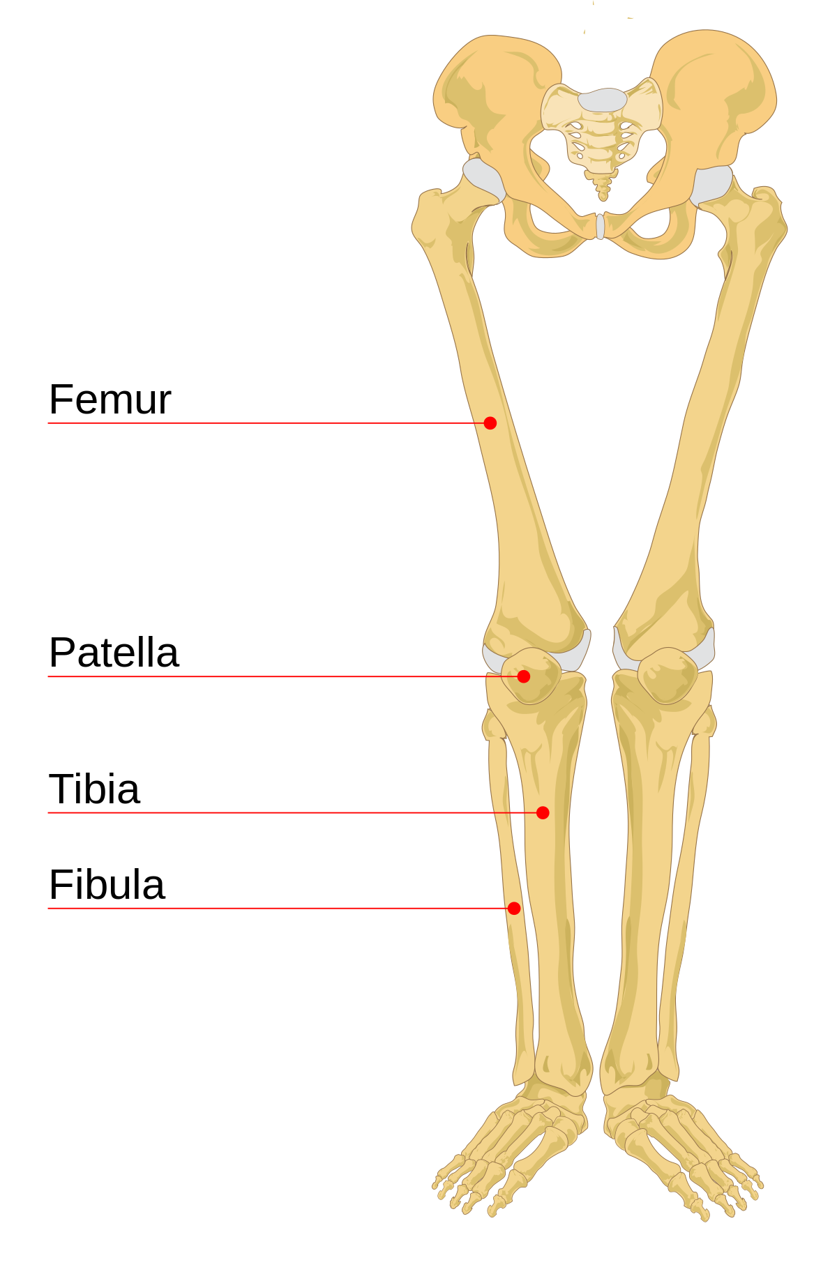
Relapse multifocal osteomyelitis secondary to septicemia: License image the bones of the leg are the femur, tibia, fibula and patella. Cheek bone (zygoma) upper jaw (maxilla). Can i also check that what you really require by arm and leg bone. Related online courses on physioplus. Learn the bones of the body with skeletal system quizzes. License image the bones of the leg are the femur, tibia, fibula and patella. In uberon this is stylopod + zeugopod but excluding autopod. Start learning with our skeleton diagrams, bone labeling exercises and skeletal system quizzes! The bones of the leg are the femur, tibia, fibula and patella. Bones of the pelvic girdle. Its lower end helps create the knee joint. In the lower leg, the tibia bears most of the body's weight while the fibula supports the muscles of balance in the lower leg and ankle.
Hip and thigh knee and leg ankle and foot neurovasculature of the lower limb leg bone diagram. The ends have red marrow.
Share :
Post a Comment
for "Right Leg Bone Diagram - Joints Ligaments And Connective Tissues Advanced Anatomy 2nd Ed"


Post a Comment for "Right Leg Bone Diagram - Joints Ligaments And Connective Tissues Advanced Anatomy 2nd Ed"